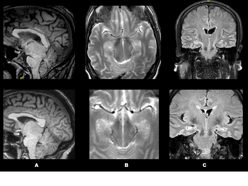

The falx cerebri seemed to develop from the tongue-like folds. Up to 9-10 weeks, the lateral mesenchymal condensations became tongue-like folds i.e., the primitive form of the tentorium cerebelli, while the membranous meninx became the diaphragma sellae. Notably, the topographical anatomy of the oculomotor, trochlear and trigeminal nerves did not change: the oculomotor nerve ran along the rostral aspect of the membranous meninx, the trigeminal nerve ran along the caudal side of the lateral mesenchymal condensation, and the trochlear nerve remained embedded in the lateral condensation. In addition, a mesencephalic or cranial flexure, has also developed at the junction between the mesencephalon and the diencephalon. Subsequently, the plica ventralis evolved into 3 parts: bilateral lateral mesenchymal condensations and a primitive membranous meninx extending between. In the loose tissues of the plica, the first meninx appeared as a narrow membrane along the oculomotor nerve at 7-8 weeks. We histologically examined paraffin-embedded sections from 18 human embryos and fetuses at 6-12 weeks of gestation. Dopamine neurons of the VTA are important for behavioral regulation in response to rewarding substances, including natural rewards and addictive drugs. A distinct mesenchymal structure, the so-called plica ventralis encephali, is sandwiched by the fetal mesencephalic flexure. Dopamineglutamate co-release is a unique property of midbrain neurons primarily located in the ventral tegmental area (VTA). Development of the meninges in and around the plica ventralis encephali has not been well documented. The first immunoreactive neurons appeared very early since they were found on day 30 of pregnancy in the medioventral part of the mesencephalic flexure.


 0 kommentar(er)
